Function Of Calcaneofibular Ligament
Java 32 bit for windows 7 Windows 7 - Free Download Windows 7 java 32 bit for windows 7 - Windows 7 Download - Free Windows7 Download. Java for windows 7 32 bit download free download - Windows 7 (Professional), Java Development Kit (32 bit), nVidia Graphics Driver (Windows Vista 32-bit / Windows 7 32-bit / Windows 8 32-bit),. Java software for your computer, or the Java Runtime Environment, is also referred to as the Java Runtime, Runtime Environment, Runtime, JRE, Java Virtual Machine, Virtual Machine, Java VM, JVM, VM, Java plug-in, Java plugin, Java add-on or Java download. Java win7 32 bit download. Both 32-bit and 64-bit browsers, you need to download both 32-bit and 64-bit Java, respectively; Verify if you are using 32-bit or 64-bit browser Internet Explorer. Launch Internet Explorer browser. Click on the Help tab at the top. Select About Internet Explorer which will bring up an information window.
- Function Of Calcaneofibular Ligament Pain
- Calcaneofibular Ligament Tear
- Function Of Talofibular Ligament
| Anterior talofibular ligament | |
|---|---|
The ligaments of the foot from the lateral aspect (anterior talofibular ligament labeled at bottom center left) | |
Lateral view of the human ankle (anterior talofibular ligament labeled at center right) | |
| Details | |
| From | Talus bone |
| To | Fibula (lateral malleolus) |
| Identifiers | |
| Latin | Ligamentum talofibulare anterius |
| TA | A03.6.10.009 |
| FMA | 44083 |
| Anatomical terminology | |
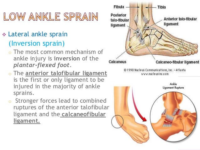
The anterior talofibular ligament is a ligament in the ankle. It passes from the anterior margin of the fibular malleolus, anteriorly and medially, to the talus bone, in front of its lateral articular facet. It is one of the lateral ligaments of the ankle and prevents the foot from sliding forward in relation to the shin. It is the most commonly injured ligament in a sprained ankle—from an inversion injury—and will allow a positive anterior drawer test of the ankle if completely torn.
- Dec 23, 2011 The main lateral (outside of ankle) ligaments involved in ankle sprains are named for the two bones they connect and are involved in ankle sprains usually in the following order 1) Anterior talofibular ligament (ATFL) (L1 in figure), 2) Calcaneofibular ligament (CFL) (L2 in figure), and rarely 3) Posterior talofibular ligament (PTFL) (L3 in.
- The anterior talofibular ligament is a ligament in the ankle. It passes from the anterior margin of the fibular malleolus, anteriorly and medially, to the talus bone, in front of its lateral articular facet. It is one of the lateral ligaments of the ankle and prevents the foot from sliding forward in relation to the shin. It is the most commonly injured ligament in a sprained ankle—from an inversion injury—and will allow a.
- Jan 23, 2014 The calcaneofibular ligament attaches to the calcaneus and the distal tip of the fibula. It stabilizes both the ankle and the subtalar joint. The posterior talofibular ligament attaches to the calcaneus and the posterior aspect of the distal fibula.
- Feb 28, 2014 Ankle joint ligament (calcaneofibular ligament) is superficial and ligament lies between skin and ankle bone. Direct impact is often associated with skin laceration and ankle joint ligament injury or tear. Causes of direct impact causing ankle joint ligament injury is as follows.
- The objective of this study was to perform a systematic literature review of the last 10 years regarding evidence for the treatment and prevention of lateral ankle sprains. Surgical and non-surgical treatment, immobilization versus functional treatment, different external supports, balance training.
Successive section of the lateral collateral ligaments was performed, including, in particular, selective division of the short and long fibres of the posterior talofibular ligament. The function of this ligament was investigated in combination with the other two collateral lateral ligaments, with the calcaneofibular ligament alone, and finally.
See also[edit]
References[edit]
This article incorporates text in the public domain from page 351 of the 20th edition of Gray's Anatomy (1918)

Further reading[edit]
- Matsui K, Takao M, Tochigi Y, Ozeki S, Glazebrook M (June 2017). 'Anatomy of anterior talofibular ligament and calcaneofibular ligament for minimally invasive surgery: a systematic review'. Knee Surgery, Sports Traumatology, Arthroscopy (Review). 25 (6): 1892–1902. doi:10.1007/s00167-016-4194-y. PMID27295109.
External links[edit]
- Anatomy figure: 17:10-04 at Human Anatomy Online, SUNY Downstate Medical Center - 'Lateral view of the ligaments of the ankle.'
- lljoints at The Anatomy Lesson by Wesley Norman (Georgetown University) (latanklejoint)
| Plantar plate | |
|---|---|
| Details | |
| From | Metatarsal and phalanx |
| To | Phalanx |
| Identifiers | |
| Latin | ligamenta plantaria |
| MeSH | D000069262 |
| TA | A03.6.10.803 |
| FMA | 71425 |
| Anatomical terminology | |
In the human foot, the plantar or volar plates (also called plantar or volar ligaments) are fibrocartilaginous structures found in the metatarsophalangeal (MTP) and interphalangeal (IP) joints. The anatomy and composition of the plantar plates are similar to the palmar plates in the metacarpophalangeal (MCP) and interphalangeal joints in the hand; the proximal origin is thin but the distal insertion is stout. Due to the weight-bearing nature of the human foot, the plantar plates are exposed to extension forces not present in the human hand.[1]
The plantar plate supports the weight of the body and restricts dorsiflexion, whilst the main collateral ligament and the accessory collateral ligament (together referred as the collateral ligament complex, CLC) prevent motions in the transverse and sagittal planes.[2]The major difference between the plantar plates of the MTP and IP joints is that they blend with the transverse metatarsal ligament in the MTP joints (not present in the toes). The MTP joint of the first toe differs from those of the other toes in that other muscles act on the joint, and in the presence of two sesamoid bones.
The plantar plate is firm but flexible fibrocartilage with a composition similar to that found in the menisci of the knee (composed roughly of 75% type-I collagen), and can thus withstand compressive loads and act as a supportive articular surface. Most of its fibers are oriented longitudinally, in the same direction as the plantar fascia, and the plate can thus sustain substantial tensile loads in this direction.[2]
Metatarsophalangeal joints[edit]
At the metatarsophalangeal joint the plantar plate plays an important role in the foot's weight-bearing function. Program has stopped working error windows 7.
The plantar plate is attached to the proximal phalanx, to the major longitudinal bands of the plantar fascia, and to the collateral ligaments. Together with the collateral ligaments, it forms a soft tissue box which is connected to the sides of the metatarsal head. The plate from the substantial distal insertion of the plantar fascia and can withstand tensile loads in line with the fascia itself. The plate can witstand compressive loads from the metatarsal head because of the orientation of the fibers in its fibrocartilage.[3]
The skeleton of the foot rests on a multi-layered ligamentous system of beams and trusses that responds to weight-bearing on irregular surfaces. A transverse system at the MTP joints is formed by the plantar plates and the deep transverse metatarsal ligament. The strong, longitudinal fibres of the deep plantar fascia are inserted along this transverse system to form a strong longitudinal system. The longitudinal system controls the longitudinal arches of the foot, whilst the transverse system controls the splay of the forefoot. Both systems are centered on the plantar plates and activated weight-bearing pressure on the metatarsal heads.[4]
The tendon of the extensor digitorum longus muscle extends the MTP joint by using the plantar fibroaponeurotic structure as a sling. The muscle becomes a deforming force if the MTP joint is held in an extended position over a long time, such as in a high-heeled footwear. The muscle extends at the IP joints when the MTP joint is flexed or in neutral position. Flexion is primarily performed by intrinsic foot muscles; the second toe (the) is unique as it has two dorsal interossei but no plantar interossei muscles. The lumbrical muscles, attached to the medial side of the lesser toes, act as unopposed adductor, but become insufficient plantar flexors with chronic extension.[5]
Function Of Calcaneofibular Ligament Pain
Notes[edit]
Calcaneofibular Ligament Tear
- ^Johnston, Smith & Daniels 1994
- ^ abMohana-Borges et al. 2003, Discussion, pp. 179–80
- ^Deland et al. 1995
- ^Stainsby 1997, pp. 60–1
- ^Canale, Beaty & Ishikawa 2008, Anatomy
References[edit]
- Canale, S. Terry; Beaty, James H.; Ishikawa, Susan N. (2008). 'Hammer Toe Correction'. Procedures Consult (Elsevier). Archived from the original on 2010-07-26. Retrieved 25 November 2010.
- Deland, JT; Lee, KT; Sobel, M; DiCarlo, EF (August 1995). 'Anatomy of the plantar plate and its attachments in the lesser metatarsal phalangeal joint'. Foot Ankle Int. 16 (8): 480–6. doi:10.1177/107110079501600804. PMID8520660.
- Johnston, RB 3rd; Smith, J; Daniels, T (May 1994). 'The plantar plate of the lesser toes: an anatomical study in human cadavers'. Foot Ankle Int. 15 (5): 276–82. doi:10.1177/107110079401500508. PMID7951967.
- Mohana-Borges, Aurea V. R.; Theumann, Nicolas H.; Pfirrmann, Christian W. A.; Chung, Christine B.; Resnick, Donald L.; Trudell, Debra J. (April 2003). 'Lesser Metatarsophalangeal Joints: Standard MR Imaging, MR Arthrography, and MR Bursography—Initial Results in 48 Cadaveric Joints'. Radiology. 227 (1): 175–182. doi:10.1148/radiol.2271020283. PMID12668744.
- Stainsby, G D (January 1997). 'Pathological anatomy and dynamic effect of the displaced plantar plate and the importance of the integrity of the plantar plate-deep transverse metatarsal ligament tie-bar'. Annals of the Royal College of Surgeons of England. 79 (1): 58–68. PMC2502622. PMID9038498.


